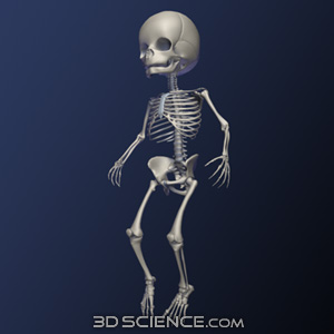
3D Infant Skeleton
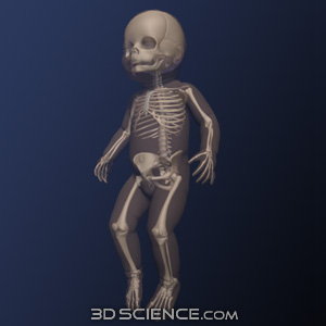
The model here shown of the Infant with skeleton is both high in detail and resolution. This model was developed by medical illustrators using both photographic and illustrative atlases to adapt the fully matured skeleton to that of a three-month-old infant, maintaining accuracy for the project at hand. Noticeable differences are seen especially in the cranial regions, where the fontanelles have yet to fully fuse together to allow growth of the infant brain. This model includes the infant skin free with the skeleton.
Zygote's Solid 3D Anatomy has set the standard in CAD and Simulation for over a decade.
- Formats:
- 3D Studio Max
- Blender (OBJ)
- Cinema 4D
- Generic OBJ
- Maya
- Polygons (as tris): 111000
- UV Coordinates: No
- Textures: No
- Grouping: Yes
- Delivery Method: Download
- Price: $2,575
- Rigged: No
"We wanted to demonstrate the kind of 3D experience we should all expect from modern browsers. We chose the human body because of how fascinating it is to learn about it, and we chose to work with Zygote Media Group because of their outstanding imagery and team."
Roni Zeiger, MD / Google's Chief Health Strategist
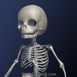
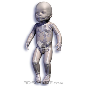
The model here shown of the Infant with skeleton is both high in detail and resolution. This model was developed by medical illustrators using both photographic and illustrative atlases to adapt the fully matured skeleton to that of a three-month-old infant, maintaining accuracy for the project at hand. Noticeable differences are seen especially in the cranial regions, where the fontanelles have yet to fully fuse together to allow growth of the infant brain. This model includes the infant skin free with the skeleton.
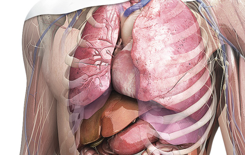
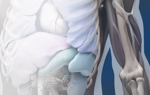
Creators of the world's leading 3D human anatomy models for use in medical illustration, animation, engineering, simulation, and anatomy software products.
2015 © All Rights Reserved. Privacy Policy | Terms of Service | Careers