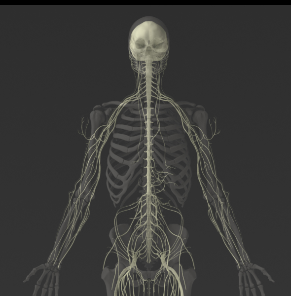
Solid 3D Male Nervous System
Need to model a system involving the Brain or Spinal Cord?
Zygote's detailed 3D Male Nervous System is perfect for:
- Head Trauma Research
- Safety Systems Studies
- Simulation of Medical Procedures
- CAD Product & Device Design
With brain and spinal cord geometries developed from medical scans, these high fidelity 3D geometries can be depended on for accurate results.
A free eDrawing of this model is available. What is an eDrawing? Click here to find out more.
Zygote's Solid 3D Anatomy has set the standard in CAD and Simulation for over a decade.
- Formats:
- ProE/Creo
- IGES
- ParaSolid
- SolidWorks
- Step
- Delivery Method: Download
- Price: $5,040
"One of the main reasons we (Ghost) upgraded was to take advantage of the improved texturing and increased accuracy of the bony anatomy. Historically, we've spent a goodly amount of time trying to massage previous versions into shaders and geometry tweaks that stood up to the scrutiny of a close-up shot. With version 5 of the male and female anatomy collections, all I did was light it. No screwing around. Just animate, light and render... Honestly? The surgical procedure portions were the easiest part of this production."
Laura Schulz / Project Manager - ghOst Productions, Inc.
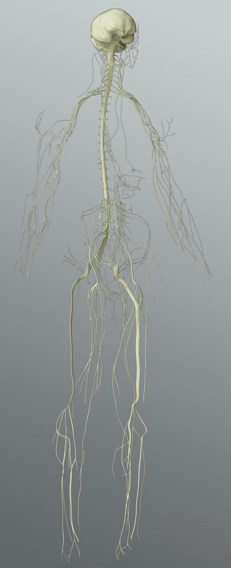
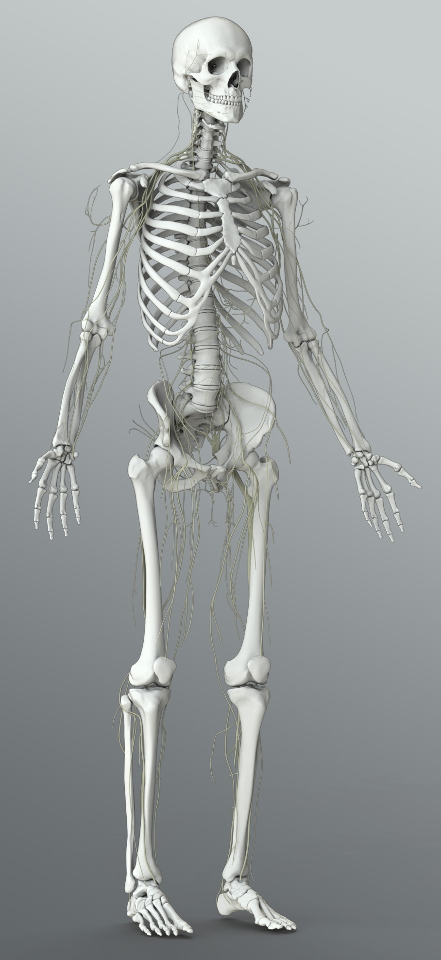
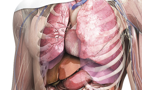
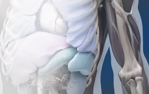
The Zygote Solid 3D Male Nervous System includes the highly detailed Brain, Meninges and Spinal cord with nerve schematic throughout the body to the arms and legs. Developed from medical scans, photographs, atlases and published references, this collection includes detail sufficient for intensive modeling in specific regions of the head and back with meninges geometries that can serve as light stand-ins for whole-system modeling.
The Zygote Solid 3D Brain is a very highly-detailed, medically-accurate model of a human brain. Created from MR scan data, the cerebral geometry accurately defines typical cerebral landmark sulci, gyri and ventricles. Also available are Solid 3D models detailing the volumes of the Arachnoid Membrane and the Dura Matter including the Falx Cerebri and Tentorum Cerebelli. The Meninges accurately accommodate the cerebral circulation (licensed separately) including the Superior Sagittal Sinus and the Transverse Sinus. The Trigeminal, Optic, and Nasal Nerves are all correctly represented from scan data. Other cranial nerves have been created using photographic and illustrative reference. The Cerebrum, Cerebellum, and Brain Stem are all one solid part. The Arachnoid Membrane and Dura Matter are each additional separate parts.
The Spinal cord was created by digitizing photographs of cross sections of dissected every inch and interpolating shape transitions from one digitized section to the next. The size, contour, posture and skeletal correlation of the Spinal cord were derived using medical scan data. The encasing Dura mater with offset spacing between Spinal cord and the Dura was developed using offsets observed in medical scan data.
The nerve tract schematic throughout the rest of the body and into the limbs were developed by medical modelers using published reference material including atlases and photographs.
Creators of the world's leading 3D human anatomy models for use in medical illustration, animation, engineering, simulation, and anatomy software products.
2015 © All Rights Reserved. Privacy Policy | Terms of Service | Careers